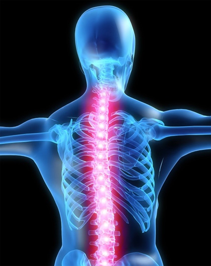
Spinal X-Rays
Spinal X-rays are also done to check the curve of your spine (scoliosis) or for spinal defects.
X-rays are a form of radiation, like light or radio waves, that are focused into a beam, much like a flashlight beam. X-rays can pass through most objects, including the human body.
Dense tissues in the body, such as bones, block (absorb) many of the X-rays and look white on an X-ray picture. Less dense tissues, such as muscles and organs, block fewer of the X-rays (more of the X-rays pass through) and look like shades of gray on an X-ray. X-rays that pass only through air look black on the picture.
- Cervical spine X-ray – This X-ray test takes pictures of the 7 neck (cervical) bones.
- Thoracic spine X-ray – This X-ray test takes pictures of the 12 chest (thoracic) bones.
- Lumbosacral spine X-ray – This X-ray test takes pictures of the 5 bones of the lower back (lumbar vertebrae) and a view of the 5 fused bones at the bottom of the spine (sacrum).
- Sacrum/coccyx X-ray – This X-ray test takes a detailed view of the 5 fused bones at the bottom of the spine (sacrum) and the 4 small bones of the tailbone (coccyx).
The most common spinal X-rays are of the cervical vertebrae (C-spine films) and lumbosacral vertebrae (LS-spine films).
- Find the cause of ongoing pain, numbness, or weakness.
- Check for arthritis of the joints between the vertebrae and the breakdown (degeneration) of the discs between the spinal bones.
- Check injuries to the spine, such as fractures or dislocations.
- Check the spine for effects from other problems, such as infections, tumors, or bone spurs.
- Check for abnormal curves of the spine, such as scoliosis, in children or young adults.
- Check the spine for problems present at birth (congenital conditions), such as spina bifida, in infants, children, or young adults.
- Check changes in the spine after spinal surgery.
Before the X-ray test, tell your doctor if you:
- Are or might be pregnant. The risk of radiation exposure to your unborn baby (fetus) must be considered. The risk of damage from the X-rays is usually very low compared with the potential benefits of the test. If a spinal X-ray is absolutely necessary, a lead apron will be placed over your belly to shield your baby from the X-rays.
- Have had an X-ray test using barium contrast material (such as a barium enema) in the past 4 days. Barium shows up on X-ray films and makes it hard to get a clear picture of the lower back (lumbar spine).
You don’t need to do anything else before you have this test.
Reviews
Shawn is absolutely awesome. I was under severe pain this Monday and Shawn came to the clinic despite being a holiday and provided necessary adjustment that helped me sleep at least and pain came down to less than 50% in the next few days. He is very helpful in accommodating changes and giving suggestions.
Shawn is absolutely awesome. I was under severe pain this Monday and Shawn came to the clinic despite being a holiday and provided necessary adjustment that helped me sleep at least and pain came down to less than 50% in the next few days. He is very helpful in accommodating changes and giving suggestions.


